Compression of the superficial peroneal nerve (SPN) is due to the superficial fascial layer that encapsulates the SPN and its distal entrapment point called the transverse crural ligament. These structures are typically the cause for numbness and pain in the territory of the SPN. Release of the SPN involves the longitudinal release of the superficial fascial layer and the transverse crural ligament. A lateral and anterior fasciotomy is also performed both longitudinally and transversely. Care is taken to look for two branches of the SPN and decompress both branches. This release is performed on patients that present with peroneal neuropathy that fail to resolve from conservative management and have symptoms that localize to the territory of the SPN. In this case, the patient presented with a complex history of neuropathic pain in the lower left leg following multiple knee surgeries over a span of many years. During examination, the patient was able to tolerate light touch related to compression-type injury rather than withdrawing from severe pain in keeping with a neurectomy-type injury; thus compression neuropathy and not neuroma injury was suspected. The scratch collapse test with ethyl chloride revealed provocation, first at the common peroneal nerve at the fibular head, then second at the saphenous nerve in the thigh, and then third at the SPN. Her surgical management included the release of these three nerves. This video outlines the surgical technique for releasing the SPN in the lower leg.
Standard:

Extended:

POSITION
Supine with tourniquet control.
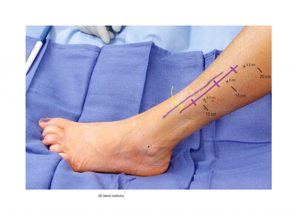
INCISION
A longitudinal incision is centered over a muscle bulge and interval between the peroneus longus and extensor digitorum longus. The incision is marked at approximately 3.5, 4, and 4.5 cm lateral to the spine of the tibia at 10, 15, and 20 cm proximal to the lateral malleolus. It can be extended as necessary for a complete release.
POST-OPERATIVE MANAGEMENT
The incision is closed with absorbable suture and glue. A soft compression dressing is applied. Patients are allowed to weight-bear as tolerated. Assistive devices are offered to patients post-operatively but typically are not needed. Patients are instructed to avoid unnecessary ambulation and elevate the leg as much as possible during the first three weeks post-operatively. This will help limit swelling of the lower extremity and any issues with wound healing. Diabetic and obese patients may require additional interrupted nylon sutures to support their incision. These are removed at two weeks post-operatively. At three weeks post-operatively, patients may be referred to physical therapy to address any issues with edema, scar, ankle ROM, strength and ambulation. At two months post-operatively, they are typically released to all activities.
REFERENCES
- Franco MJ, Phillips BZ, Lalchandani GR, Mackinnon SE. Decompression of the superficial peroneal nerve: clinical outcomes and anatomical study. J Neurosurg. 2016 Apr 22:1-6. PMID: 27104849.
- Humphreys DB, Novak CB, Mackinnon SE. Patient outcome after common peroneal nerve decompression. J Neurosurg. 2007 Aug;107(2):314-8. PMID: 17695385.


Disclosure: No authors have a financial interest in any of the products, devices, or drugs mentioned in this production or publication.