Compression of the common peroneal nerve at the fibular head is under recognized. Symptoms range from numbness and tingling in the posterior and lateral aspect of the lower leg and dorsal aspect of the foot to foot drop in severe cases of denervation. Symptoms can be exaggerated when the peroneal nerve is under tension during knee extension. The compression point of the common peroneal nerve is just distal to the fibular head as it dives deep to the peroneus longus. This is where the posterior crural intermuscular septum deep to the peroneus longus is found to be the primary contributing factor for the compression symptoms. Release of the common peroneal nerve involves dividing this intermuscular septum in addition to other tendinous septum planes and the tendinous fascia deep to the peroneal nerve.
Standard:

Extended:

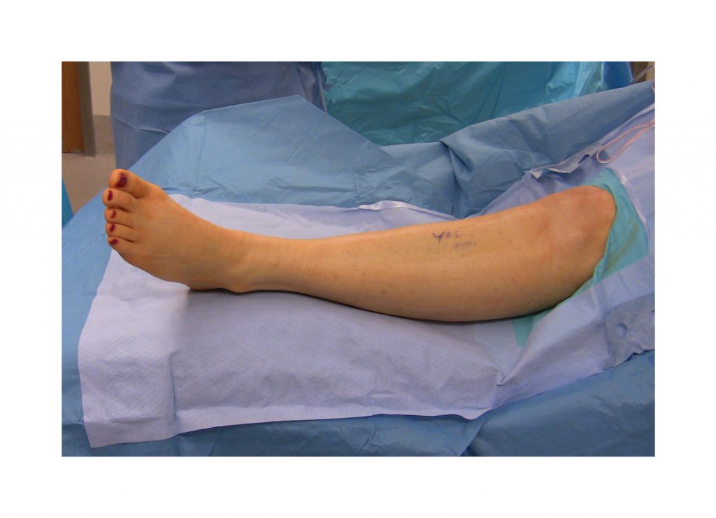
POSITION
Supine with foot propped on a sand bag in keeping with knee flexion at around 45. Tourniquet control is preferred for optimal visualization.
INCISION
An oblique incision is made just inferior to the fibular head and oriented from posterior-proximal to anterior-distal along the course of the common peroneal nerve.
POST-OPERATIVE MANAGEMENT
The incision is closed with absorbable suture and glue. A soft compression dressing is applied. Patients are allowed to weight-bear as tolerated. Assistive devices are offered to patients post-operatively but typically are not needed. Patients are instructed to avoid unnecessary ambulation and elevate the leg as much as possible during the first three weeks post-operatively. This will help limit swelling of the lower extremity and any issues with wound healing. Diabetic and obese patients may require additional interrupted nylon sutures to support their incision. These are removed at two weeks post-operatively. At three weeks post-operatively, patients may be referred to physical therapy to address any issues with edema, scar, ankle ROM, strength and ambulation. At two months post-operatively, they are typically released to all activities.
REFERENCES
- Humphreys DB, Novak CB, Mackinnon SE. Patient outcome after common peroneal nerve decompression. J Neurosurg. 2007 Aug;107(2):314-8. PMID: 17695385.


Disclosure: No authors have a financial interest in any of the products, devices, or drugs mentioned in this production or publication.